Nina Sarvašová, Michaela Gajdošová, Jakub Dvořák, František Štěpánek
Magnetic Resonance Imaging (MRI) is the non-destructive and non-invasive imaging technique based on the principles of nuclear magnetic resonance (NMR), however, it is not to be confused with the spectroscopic method NMR used in scientific laboratories to obtain the physical and chemical information about the molecules. The human body primarily consists of fat and water, both of which contain hydrogen atoms and thus make human body by approximately 63% hydrogen atoms. Atom nuclei having non-zero net magnetic spin (odd number of nucleons) produce NMR signal and H1 with its natural abundance of more than 99% and single proton in the nuclei belongs to such atoms. Therefore, MRI images mostly the NMR signal from hydrogen nuclei.
Each voxel of an image of the human body contains one of more organic tissues containing hydrogen in one of its forms. These hydrogen atoms can absorb and emit the radio frequency (RF) energy in the range of the electromagnetic spectrum and thus produce NMR signal dependent on the number of atoms with non-zero net spin contained within the voxel. The spatial distribution of the signal is obtained by modulating the magnetic field using gradients, which results in the variations in the phase and frequency of the RF energy absorbed and emitted by the voxels contained in the scanned area.
The MRI scanners are primarily used for medical purposes, however they also provide for great opportunities within the scientific research. The Laboratory of Chemical Robotics dispose of the MRI ICON Desktop system (BRUKER, magnetic field strength 1T, gradients 450mT/m). It allows for various kinds of imaging, i.e. solid forms dissolution, particle adhesion, 3D structure imaging and so on, and can be also used in flow setting when connected to the outer flow circuit with peristaltic pump (Fig. 1, 2, 3).
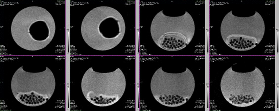
Figure 1: The dissolution of the gelatin caffeine tablet.
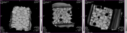
Figure 2: The inner bioreactor matrix overgrown with fungus.
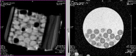
Figure 3: Alginate microparticles containing Au nanoparticles: proton density weighted scan (left), T2 weighted scan (right).
Publications
- Podgórna K., Szczepanowicz K., Piotrowski M., Gajdošová M., Štěpánek F., Warszyński P., “Gadolinium alginate nanogels for theranostic applications”, Coll. Surf. B 153, 183-189 (2017)
- Gajdošová M., Pěček D., Sarvašová N., Grof Z., Štěpánek F., “Effect of hydrophobic inclusions on polymer swelling kinetics studied by magnetic resonance imaging”, Int. J. Pharm. 500, 136-143 (2016)
- Sarvašová N., Ulbrich P., Tokárová V., Zadražil A., Štěpánek F., “Artificial swarming: towards radiofrequency control of reversible micro-particle aggregation and deposition”, Powder Technol. 278, 17-25 (2015)