Monika Majerská, Anna Krejčí, Ondřej Rychecký, František Štěpánek
Current biochemical studies of cellular physiology and behavior are mostly based on 2D cell cultures on well plates. However, these 2D cell cultures do not recreate properly in vivo systems in terms of cellular communication, gene and protein expression patterns and diffusion of soluble molecules needed for growth (e.g. respiratory gases, nutrients, growth factors etc.). The opposite end of experimental systems, being represented by animal models, is very costly, time-consuming, not always representative and raises ethical questions.
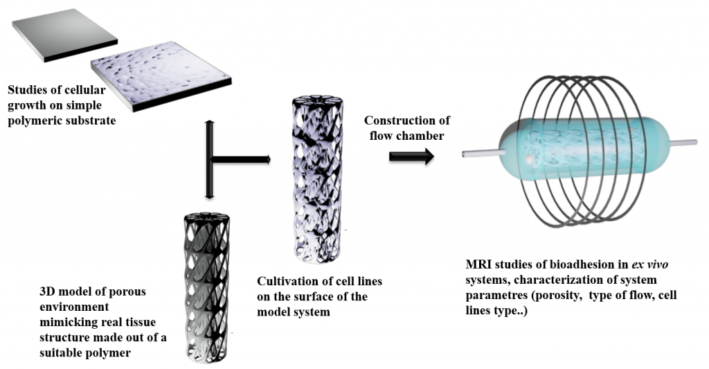
As a result, 3D cultures are currently being developed to provide the third dimension, which is essential to integrate mechanical and chemical signals, better mimicking the in vivo microenvironment and better reflecting cellular behavior. In our case, 3D cell cultures will be used to monitor the adhesion of so called chemical robots in ex vivo systems. This approach consist of mastering several parts: scaffold material, structure fabrication, inoculating suitable cells and biomolecules, cellular growth and proliferation, and finally structural and functional analysis of the formed 3D cell cultures as shown in the Figures below. Scaffold fabrication methods involve 3D printing, gas foaming and particle templating method
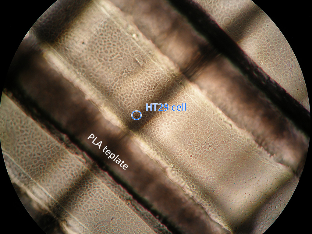
Growth of HT29 cell line on a PLA template from 3D printer
Publications
- Rychecký O., Majerská M., Král V., Štěpánek F., Čejková J., “Spheroid cultivation of HT-29 carcinoma cell line in liquid marbles”, Chem. Pap. 71, 1055-1063 (2017)